You must be signed in to read the rest of this article.
Registration on CDEWorld is free. You may also login to CDEWorld with your DentalAegis.com account.
THE CONCEPT OF BALANCE
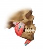
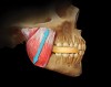
The equilibrium of the entire masticatory system is dependent on balance.1 The mandible at rest is balanced between the resting lengths of the elevator muscles and the depressor muscles (Figure 1). Anything that affects the resting length of either group of opposing muscles can affect the critical relationship of the mandible with the maxilla at the resting position. Because the teeth are not in contact at the rest position and the mandible-to-maxilla relationship is not consistent,2,3 the rest position is not an accurate determinant of the jaw-to-jaw relationship at maximum intercuspation.
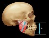
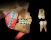
The correct determinant for the mandible-to-maxilla relationship at maximum tooth contact is a result of the repetitive contracted length of the elevator muscles (Figure 2).4 To understand this concept, the dentist must recognize the role balance plays in the eruptive process of the lower teeth toward the erupting upper teeth. Teeth erupt toward their opposing teeth until there is a balance of opposing eruptive forces (Figure 3). The stopping point for eruption occurs as the mandible is positioned vertically in relationship with the maxilla by the repetitious power cycle of the closing musculature. This relationship should be in harmony with the maximum intercuspation of the teeth during complete closure. The key to understanding the vertical dimension of occlusion (VDO) is recognition that the teeth adapt to the vertically positioned mandible as dictated by the musculature, not vice versa. If the repetitive muscle contraction length is impeded, the teeth must adapt to conform to the closing power cycle as muscle regains its optimal contraction length. An adaptive process appears to maintain the VDO throughout life through changes in the dimension of the alveolar processes as needed. Ricketts et al5 showed that lower facial height in adults stayed constant with age. McAndrews6 demonstrated in more than 1,000 orthodontic patients that changes in VDO reverted back to the original dimensions within 1 year, and this occurred whether the VDO was increased or decreased (oral ommunication, 2001).
McAndrews’ observations changed the way the dental profession looks at VDO as a factor in treatment planning because from that study dentists learned that VDO could be opened or closed without doing harm as long as they combined the change with simultaneous equal intensity contacts in centric relation (CR). If that is done, the relationship between the cementoenamel junctions of the teeth and the interceptal bone remains unchanged as the VDO increases or decreases to its original dimension in harmony with the masticatory musculature. This finding indicates that changes in VDO occur from either elongation or regressive remodeling of the dentoalveolar process.7-10 To say it another way, the alveolar bone moves with the teeth to increase or decrease the VDO as needed to conform to the consistent muscle contraction length. Since becoming aware of this phenomenon, the author has observed it consistently in hundreds of patients in which the VDO was changed. Today, clinicians recognize that they can gain some predictable advantages by changing the VDO when it is indicated specifically. Clinicians also recognize that unnecessary increases in VDO are considered overtreatment because the increased VDO cannot be maintained. Before that statement can be accepted, however, dentists must be aware of how condyle position affects VDO.
HOW CONDYLE POSITION AFFECTS VDO
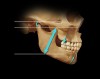
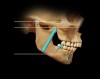
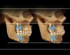
The dimension that determines VDO is located at the elevator muscles because it is the repetitive contracted length of the muscles that establishes the jaw-to-jaw vertical position at maximum intercuspation. This dimension at maximum intercuspation is controlled by the muscles, regardless of condylar position.This means that if condyles are displaced vertically down the eminentia at maximum intercuspation (Figure 4), the dimension from muscle origin to insertion is shortened when the condyles are seated up into CR (Figure 5).
This observation on mounted diagnostic casts often points to a solution for severe anterior wear if a slide from CR to a forward jaw position is responsible for the wear, a common occurrence. Seating the condyles back and up into CR allows more room to restore anterior teeth with predictable stability of the result because VDO at the anterior teeth can be increased without interference to contracted muscle length.
CLOSING THE VDO
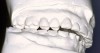
Based on McAndrews study6 and the author’s observation of hundreds of patients over a long time frame, it does not appear that there are any adverse effects from closing the VDO. The exception, however, is an important one: If closing the VDO in a deep overbite results in horizontal forces directed from the lower anterior teeth to the lingual surfaces of the upper anterior teeth, it also can result in fremitus, wear, or movement of the upper anterior teeth. Remember: the arc of closure creates a forward movement of the lower arch (Figure 6). It is this horizontal movement of lower incisal edges that sometimes can present an advantage in treatment planning for anterior teeth. Closing VDO may move the lower incisal edges forward into a better relationship for establishing stable anterior contact in CR (Figure 7A through Figure 7C).
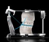
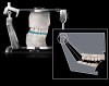
Opening the VDO moves the lower edges back, a decided advantage in some extreme wear problems in which wear has produced a near end-to-end incisal contact. In the analysis and treatment planning for many problem occlusions, the goal of anterior contact is often the determining factor that establishes the most advantageous VDO. There is only one predictable way for making decisions about the effects of altering the VDO: on articulated casts mounted in CR with a face-bow. If the condylar axis is recorded, the exact effect of changes to the VDO can be evaluated on both the anterior and posterior teeth.
EFFECT OF CHANGED VDO ON POSTERIOR TEETH
One of the most important analyses in treatment planning is observation of changes in posterior tooth contacts that are affected by changes in the VDO. A critical effect that often is ignored is the change in posterior buccolingual relationships of lower teeth with upper teeth as the wider part of the mandibular arch arcs forward to the narrower part of the maxillary arch during closure. This phenomenon occurs in almost every occlusal analysis in which gross occlusal interferences are present.
As the casts are equilibrated to allow complete closure to CR, there can be a significant forward arcing of the lower arch during closure, accompanied by a significant change in buccolingual relationships of posterior teeth. This change is in addition to the anterior–posterior relationships that change as occlusal interferences to the arc of closure are eliminated. Having seen in so many cases how critical such observations are to occlusal analysis, the author finds it hard to imagine why any dentist resists the advantages of mounted casts.
It is by observing the correct arc of opening or closing that many decisions regarding VDO can be made. Many times slight changes in VDO can eliminate arch-to-arch disharmonies on either anterior or posterior teeth, or both. If posterior teeth can benefit from restorative treatment, the decision is more acceptable. In dentitions that do not need restorative treatment, the changes may be better made via orthodontics or, in extreme cases, surgery.
THE FALLACY OF “COMFORT” AS A DETERMINANT OF VDO
One of the most prevalent misconceptions regarding VDO is the concept that trial changes can be “tested” by a response of comfort. The practice of using trial occlusal splints or provisional restorations to see if patients can tolerate the change is an unnecessary and misleading method to determine VDO. VDO is unrelated to comfort, and patients can be just as comfortable with an increase or decrease in VDO, or the same VDO they had, as long as occlusal interferences are eliminated to CR and proper excursions are perfected. This is why altering the VDO to achieve a more perfected occlusion can be so successful. Add to this understanding the observation that the muscles will cause the altered VDO to adapt back to the optimum repetitive contracted length without discomfort. In fact, patients are rarely aware of the changes that take place as the dentoalveolar process is shortening or lengthening. As the VDO returns to its original position, some minor occlusal corrections may be needed, but rarely do they cause any awareness in patients that is problematic for the dentist.
If changes in VDO caused discomfort, immediate relief would not be so common when properly made interocclusal appliances are placed for correctly diagnosed occlusomuscle disorders. Nor would predictably successful results be observed routinely when the occlusal appliance at the increased VDO is removed and the occlusion is equilibrated directly.
The key to success in these patients is not related to VDO, but rather the elimination of deflective interferences to CR and excursions. Dentists who are relying solely on occlusal appliances for relief of occlusomuscle pain would be well advised to direct their energies at learning the fine points of predictable occlusal correction as well as differential diagnosis of temporomandibular disorders (TMD), so they will know which types of TMD favorably respond to occlusal correction.
SEGMENTAL APPLIANCES
One of the great disasters in dental treatment was the use of posterior (segmental) bite-raising appliances for treating TMD. The purported rationale for the treatment was an erroneous belief that such appliances “unloaded the joints.” While it is possible to reduce the load on the temporomandibular joints (TMJs) by providing posterior occlusal contacts, it is not possible to unload the joints (prefix un meaning “no”). This is because all elevator muscles are located behind the molars and between the teeth and the TMJs. Sicher11 stated that it is a law of joint physiology that all joints are always loaded because muscle pulls across joints. An increase of occlusal height opens the VDO by rotation of the condyles and/or protrusion forward and downward, but always against the slopes of the eminentia, never by distraction away from their sockets. The other problem with posterior bite appliances is that they interfere with the repetitive length of the elevator muscles, which, in turn, intrudes the teeth that are covered by the appliance. This produces a stepped occlusion (Figure 8A and Figure 8B) along with problems of anterior instability. As anterior teeth are separated from contact, the lips have a tendency to press the upper anterior teeth lingually because there is no resistance from lower tooth contact.
“LOST” VERTICAL FROM WEAR
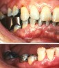
It may be difficult to change some opinions about “lost” vertical but some excellent studies refute the idea that even severe wear results in a loss of VDO.7-10 By observing the massive elongation of the alveolar process in areas of severe wear, it becomes apparent that there is a compensatory adaptation to the original VDO. This occurs even on severe habitual bruxers who wear down the teeth to the level of the bone because the bone enlarges to keep pace with the wear. Increasing the VDO on severe wear patients may be the only logical alternative to treatment because there is insufficient room to reduce tooth structure enough for the required restoration thickness. With normal alveolar processes the change in VDO will adapt back to comply12,13 with muscle lengths. The process may be aided by increases in muscle contraction force initiated by the increase in VDO.7,9,10,14 These studies showing that vertical facial dimension essentially is unaffected by even severe abrasion of the dentition demonstrated how elongation of the dentoalveolar process matches the lost vertical of the abraded teeth (Figure 9).
CONCLUSION
The mandible-to-maxilla vertical relationship at which the lower arch contacts the upper arch is established by muscle. Teeth erupt into the space between the mandible and maxilla until they contact in harmony with the repetitive contracted length of the elevator muscles. It is permissible, and often advantageous, to alter the VDO to achieve a more stable relationship of lower teeth with upper teeth because changes in VDO position the lower teeth horizontally as well as vertically on their opening/closing arc.
Changes in VDO self-adapt to the original VDO without harm or discomfort if the occlusal contacts are in harmony with CR. If the VDO must be altered, the most conservative treatment necessary to achieve an optimal esthetic and functional result should be performed.
REFERENCES
1. Dawson PE. Functional Occlusion: From TMJ to Smile Design. St. Louis, MO:Mosby Elsevier; 2006:114-129.
2. Williamson EH, Marshall DE Jr. Myomonitor rest position in the presence and absence of stress. Facial Orthop Temporomandibular Arthrol. 1986;3(2):14-17.
3. Atwood DA. A critique of research of rest position of the mandible. J Prosthet Dent. 1966;16(5):848-854.
4. Goldspink G. The Adaptation of Muscle to a New Functional Length. United Kingdom: John Wright and Sons Ltd; 1976.
5. Ricketts RM, Roth RH, Chaconas SJ, et al. Orthodontic Diagnosis and Planning: Their Roles in Preventive and Rehabilitative Dentistry. Denver, CO: Rocky Mountain Data Systems; 1982;1:15-147.
6. McAndrews I. Presentation at: The Florida Prosthodontic Seminar; 1984;Miami, FL.
7. Hylander WL. Morphological changes in human teeth and jaws in a high attrition environment. In: Dahlbert AA, ed. Orofacial Growth and Development. New York, NY:Walter De Gruyter Inc; 1977.
8. Ramfjord SP, Blankenship JR. Increased occlusal vertical dimension in adult monkeys. J Prosthet Dent. 1981;45(1):74-83.
9. Berry DC, Poole DF. Attrition: possible mechanisms of compensation. J Oral Rehabil. 1976;3(3):201-206.
10. Crothers A, Sandham A.Vertical height differences in subjects with severe dental wear. Euro J Orthod. 1993;15(6):519-525.
11. Sicher H. The temporomandibular joint. In: Sarnat BG, ed. The Temporomandibular Joint: A Biological Basis for Clinical Practice. 2nd ed. Springfield, IL: Charles C. Thomas, Publisher; 1964.
12. Manns A,Miralles R, Palazzi C. EMG, bite force, and elongation of the masseter muscle under isometric voluntary contractions and variations of vertical dimension. J Prosthet Dent. 1979;42(6):674-682.
13. Marimoto T, Abekura H, Tokuyama H, et al. Alteration in the bite force and EMG activity with changes in the vertical dimension of edentulous subjects. J Oral Rehabil. 1996;23(5):336-341.
14. Varrella TM, Paurio K., Wouters FR, et al. The relation between tooth eruption and alveolar crest height in a human skeletal sample. Arch Oral Biol. 1995;40(3):175-180.
About the Author
PETER E. DAWSON, DDS
Founder, The Dawson Academy
St . Petersburg, Florida